Cases

Incredible results and inspiring journeys – see some of our patients’ before and after photos next time you visit our practice.

FREE GINGIVAL GRAFT #24

Pre-op

2-month post-op

Picture2

• Increase keratinized gingiva
• Improved gingival biotype (gingival thickness)
• Removal of aberrant frenum
• Slight reduction in recession
• Better plaque control and oral hygiene

• Lack of keratinized gingiva
• Aberrant frenal position
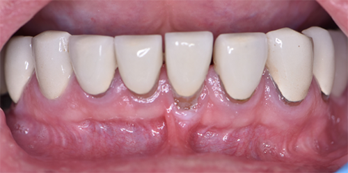
• Recession #24 – Miller: Class III recession defect & RT2
• Interproximal loss of attachment
• Discomfort while brushing
• Poor plaque control

Subepithelial Connective Tissue Graft #12

Pre-op

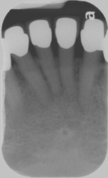
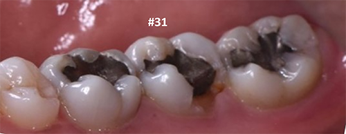
• Patient complained of sensitivity around #12
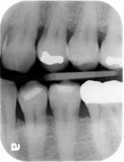
• Gingival Recession #12- Miller Class III and RT2 for #12 (interproximal attachment and bone loss around #12)
• 100% root coverage cannot be anticipated because of attachment and bone loss

6-month post-op

6Month-Post-op

• Reduction in root sensitivity around #12
• Better plaque control
• Some root coverage achieved around #12

FUNCTIONAL CROWN LENGTHENING #31

Pre-op

Deep carious lesion on lingual of #31 with invasion of biologic width (supracrestal fibers) & presence of inflammation around the area #31

6-week post-op

6Month-Post-op-31

• Functional crown lengthening
(osseous resection + removal of soft tissue) was performed with establishment of new biologic width (supracrestal fibers)
• Facilitating restorative management by restorative dentist

ESTHETIC CROWN LENTHENING

Pre-op

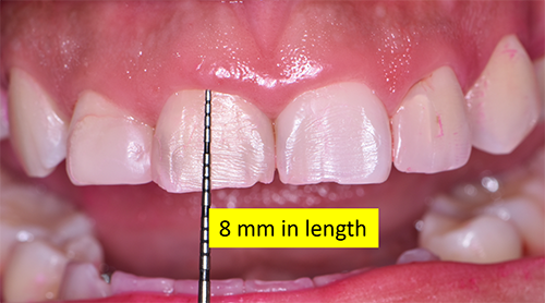
• Patient concerned with the short appearance of maxillary anterior teeth
• Minor incisal chipping (#8,9)
• Excessive gingival display on smiling
• Increased tendency of secondary caries and restorations in maxillary anterior-excess gingival display hampering oral hygiene

3-month post-op

3Month-Post-op-10mm

• Increase in length of clinical crowns
• Excessive gingival display was corrected- gingivectomy+ ostectomy & osteoplasty
• Better plaque control in maxillary anteriors
• Patient followed for restorative management

BIOPSY- Pre-op

radiograph3

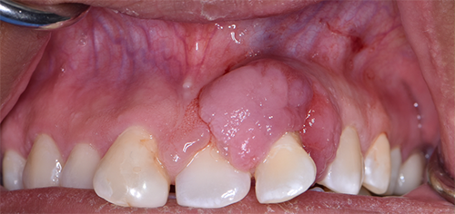
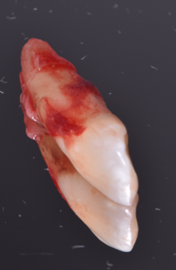
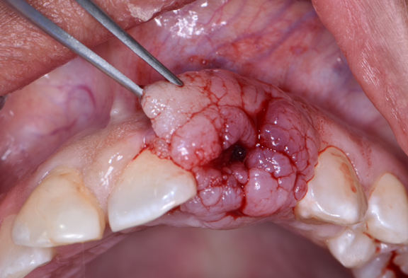
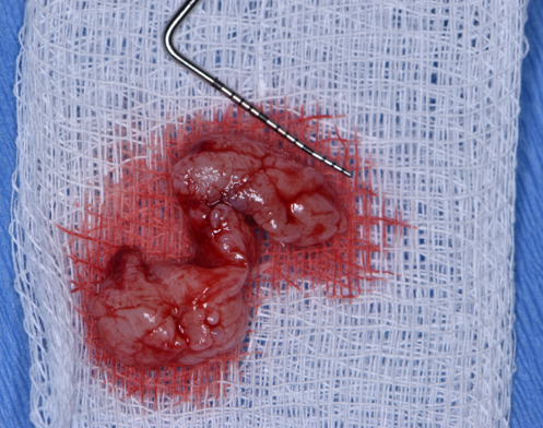
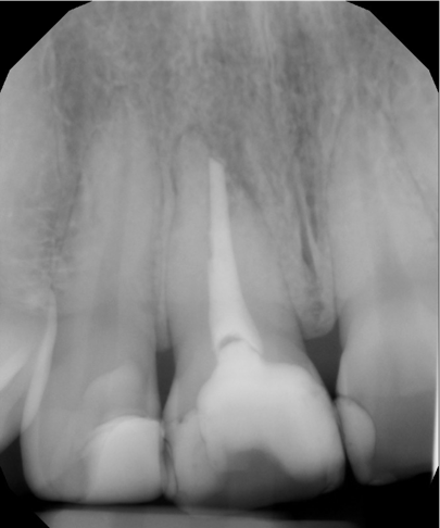
• Patient was concerned about a lesion on #10 that recently increased in size
• A well-defined, lobulated , pedunculated, pink-colored, 12x10mm lesion on the buccal surface of #10 and extending to the palatal surface.
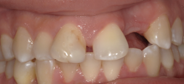
• Tooth #10 was Grade III mobile and #9 was Grade I mobile
• D/d: Pyogenic granuloma, Peripheral ossifying fibroma, Peripheral giant cell granuloma, Squamous cell carcinoma

BIOPSY- Pre-op

Lesion was removed and tooth was extracted (hopeless prognosis for the tooth) and were submitted for histopathologic examination

1-month post-op
Patient will be followed for restorative management for #10

BIOPSY- Pre-op